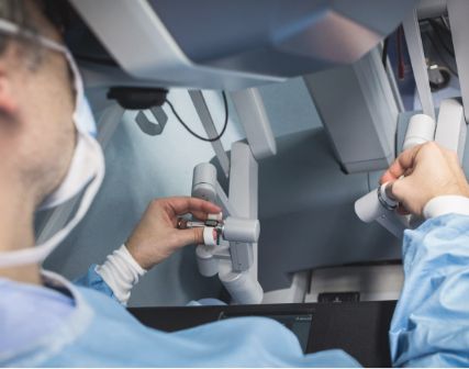
Enhancing surgeons’ education and training – Work Package 3
As part of the RHU Digital Urology 3D programme, Work Package 3 focuses on developing 3D-printed kidney model techniques for surgical training. The objective is to create biomimetic kidney models that closely replicate the anatomical and biomechanical characteristics of the human kidney, allowing surgeons to train for robot-assisted procedures and complex surgeries.
Characterising and reproducing biomechanical properties of the human kidney
The first phase of WP3 involves the MECHANICS study (UroCCR no. 115), which aims to determine mechanical characteristics of the kidney and its tumours, and to produce organ parametric maps for 3D printing.
In parallel, research will focus on developing a 3D printer and biomimetic synthetic materials—simulating human tissue—to replicate the characterised kidney.
A new study – VALIDATION (UroCCR no. 116)—will assess the realism of these 3D printed models as well as the benefit to the surgeon of training on a patient-specific 3D model prior to surgery.
Evaluating the impact on surgical training
Two additional studies will be conducted by surgeons at Bordeaux University Hospital Centre to assess the impact of 3D models on surgical training. These studies will evaluate surgeons’ perception when using 3D-printed models during simulation exercises, and compare the quality of training with and without the use of these models during the simulation.